Main Branch :Dusit Thani Commercial Complex, Muroor Road, Opposite Al Jazeera Club Abu Dhabi
At Yas Healthcare, our Radiology Department offers advanced medical imaging services to support accurate diagnosis and effective treatment. Using modern technology and patient-centered care, our team provides reliable and timely imaging for a wide range of medical conditions.
Our department operates with a fully digital PACS (Picture Archiving and Communication System), allowing imaging studies to be securely stored and instantly accessible to radiologists and referring physicians across the center. This ensures faster reporting, improved collaboration between specialists, and more efficient patient care.
We are committed to delivering high-quality, cost-effective diagnostic imaging in a comfortable and professional environment.
At Yas Healthcare, our Radiology Department offers advanced diagnostic imaging designed for accuracy, speed, and patient comfort. Using state-of-the-art technology and fully digital systems, our specialists provide reliable imaging to support precise diagnosis and effective treatment planning.
🩻 X-Ray
🖥 Ultrasound
🎗 Mammography
🦴 Bone Densitometry (DXA)
🦷 Digital Dental Panoramic Imaging
⚡ 64-Slice CT Scan (MDCT)
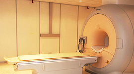
🧠 1.5T MRI
Our team is committed to delivering high-quality, efficient, and patient-focused radiology services for the Yas Healthcare community.
No special preparation is needed.
Bone densitometry, also known as a DEXA scan, is a quick and non-invasive test used to measure bone density and detect osteoporosis, a condition that weakens bones and increases the risk of fractures, especially in the hip and spine.
Osteoporosis is most common in post-menopausal women due to decreased estrogen levels. Other risk factors include family history, early menopause, smoking, long-term steroid use, certain medications, and a small or thin body frame.
A DEXA scan measures the calcium content and strength of your bones and compares your results with others of the same age and gender. The procedure usually takes about 20 minutes, and the scan itself only takes a few seconds for each area (typically the hip and spine).
Early detection helps physicians recommend treatments or lifestyle changes to prevent fractures and maintain strong bones.
Before your DEXA scan, please follow these guidelines:
Do not take calcium supplements within 24 hours before your exam
Avoid antacid tablets or liquids (such as Tums) 24 hours before the test
Wear comfortable clothing without metal, buttons, or buckles around the abdomen, pelvis, or hip area
Inform your doctor if you recently had a barium exam or CT scan with contrast, as you may need to wait 10–14 days before your bone density test
Is the test painful?
No. A DEXA scan is completely painless and does not require injections or medication.
How long does the exam take?
The entire procedure takes about 20 minutes, with the actual scan lasting only a few seconds per area.
Is the test safe?
Yes. Bone densitometry uses a very small amount of radiation, making it a safe and commonly performed diagnostic test.
⚠ Important: If you are pregnant or think you may be pregnant, please inform your physician before undergoing any imaging test that uses X-rays.
At Yas Healthcare, our Breast Imaging Unit provides advanced diagnostic tools for the early detection and evaluation of breast conditions, including breast cancer. All mammography examinations are performed by female mammography specialists, ensuring a comfortable and supportive environment for our patients. Most screening mammograms take about 15 minutes.
A mammogram is a specialized X-ray of the breast used to detect early signs of breast cancer, including microcalcifications and breast masses. Early detection allows for faster diagnosis and more treatment options.
At Yas Healthcare, we use the MammoPad, a soft foam cushion placed between the breast and the mammography machine. This technology helps make the exam warmer, gentler, and more comfortable.
Breast ultrasound is often used alongside mammography, especially for women with dense breast tissue. It helps evaluate breast lumps and determine whether they are solid or fluid-filled cysts.
Using Color Doppler and Elastography, ultrasound provides a more detailed and comprehensive assessment of breast abnormalities.
If an area appears suspicious on imaging, a breast biopsy may be recommended. During this procedure, a small sample of tissue is removed and examined in a laboratory. A biopsy is the only definitive way to determine whether a breast abnormality is cancerous.
For women at higher risk of breast cancer, or when additional imaging is required, Breast MRI may be recommended. This advanced imaging technique provides highly detailed images to support accurate diagnosis and monitoring.
Our radiologists carefully analyze each exam and compare it with previous studies whenever available to ensure the most accurate interpretation.
Women are encouraged to begin monthly breast self-examinations from the age of 20.
The best time to perform a self-exam is one week after your menstrual period, or on the same day each month if you no longer have periods.
Regular self-exams help you become familiar with your breasts so you can quickly notice any changes, lumps, or unusual symptoms. If you notice anything unusual, consult your doctor promptly.
Early detection remains the best defense against breast cancer.
Most guidelines recommend starting screening at age 40. Women at average risk should have a mammogram every two years, while those at higher risk may need annual mammograms and breast MRI.
Compression spreads the breast tissue so the radiologist can obtain clearer images and detect abnormalities more accurately.
Some deodorants and powders contain metallic particles that may appear on mammogram images and interfere with the results.
Caffeine may make the breasts more sensitive or tender, which can make the examination slightly uncomfortable.
Mammography is the most effective screening tool for early breast cancer detection.
Early detection significantly improves treatment outcomes and survival rates.
Screening mammography helps identify cancer before symptoms appear.
Some women may require additional tests if results are inconclusive.
Comfort during the exam can be improved by scheduling the mammogram between days 7 and 12 of the menstrual cycle.
At Yas Healthcare, we are committed to providing advanced breast imaging technology, compassionate care, and accurate diagnostics to support women’s health.
At Yas Healthcare, we are proud to offer advanced imaging with our 64-slice Multidetector Computed Tomography (MDCT) scanner – Philips Brilliance. This high-end technology allows us to perform a wide range of advanced CT examinations with high image quality, faster scanning times, and lower radiation exposure compared to older CT systems.
Our CT services include specialized examinations such as:
High-Resolution CT (HRCT) of the lungs
Coronary CT Angiography (CTA)
CT Perfusion Studies
CT Enterography (CTE)
Dynamic Contrast-Enhanced CT for liver evaluation
Routine CT scans of the brain, chest, abdomen, spine, and other body areas
A CT scan uses X-rays and advanced computer technology to create detailed cross-sectional images of the body. Unlike standard X-rays, which produce a flat image, CT scans generate multiple thin slices that can be reconstructed into high-resolution 3D images.
During the exam, the patient lies on a scanning table that moves slowly through the CT scanner while the X-ray tube rotates around the body. These images allow physicians to see detailed internal structures and diagnose a wide range of medical conditions.
You can usually wear your normal comfortable clothing, but we recommend avoiding items containing metal such as:
Under-wired bras
Clothing with zippers or metal buttons
Jewelry or accessories
In some cases, you may be asked to change into a hospital gown to ensure the best imaging results.
Most CT examinations take 5–15 minutes once the scanning begins.
For some scans, you may be asked to drink a special contrast liquid 30–60 minutes before the procedure to help improve image clarity.
You may bring someone with you if you wish, but it is not required. Comfortable waiting areas are available for accompanying visitors.
Stay well hydrated before your exam whenever possible.
Some CT scans require fasting (no solid food) for 2–4 hours before the procedure.
For certain exams, you may be asked to drink water or oral contrast to improve visualization of the stomach and intestines.
Detailed preparation instructions will be included in your appointment confirmation.
Patients taking Metformin (commonly used for Type 2 diabetes or PCOS) who have severe kidney disease may need to stop the medication 24 hours before and 48 hours after a CT scan requiring contrast. Please contact the department if you have any questions.
Some CT scans require an iodine-based contrast injection to improve visualization of organs and blood vessels.
The injection is given through a small IV line in your arm. During the injection, you may briefly experience:
A warm sensation throughout the body
A metallic taste in your mouth
A temporary feeling of needing to urinate
These sensations are normal and usually pass quickly. The contrast material is not radioactive and will naturally leave your body through the urine.
After the scan, our radiologists carefully review and interpret the images. A detailed report is then sent to the physician who requested your examination.
Results are typically available within 1–2 working days.
At Yas Healthcare, we are committed to providing accurate diagnostics, advanced imaging technology, and patient-focused care to support the best possible medical outcomes.
At Yas Healthcare, we offer advanced Cardiac CT scans using our Philips Brilliance 64-slice Multidetector CT (MDCT) system. This state-of-the-art technology allows our specialists to perform detailed heart scans, including coronary calcium scoring, CT coronary angiography (CTA), and cardiac function evaluation.
These non-invasive tests help detect coronary artery disease, evaluate heart health, and identify potential risks before symptoms appear.
All cardiac CT studies are performed under the joint supervision of experienced radiologists and cardiologists, ensuring accurate diagnosis and personalized patient care.
A cardiac CT scan is a specialized imaging test that uses advanced CT technology to produce highly detailed pictures of the heart and coronary arteries.
Unlike traditional tests, a cardiac CT scan can detect:
Narrowing or blockage in coronary arteries
Calcium deposits in artery walls
Early signs of coronary artery disease
Structural abnormalities of the heart
This information helps doctors determine the risk of heart attack and guide treatment decisions.
A coronary calcium score test measures the amount of calcium buildup inside the arteries that supply blood to the heart.
Calcium deposits indicate atherosclerosis (plaque buildup), which may increase the risk of heart disease.
Your results are compared with people of the same age and gender, helping physicians evaluate your personalized heart disease risk.
Early detection of coronary artery disease
Personalized heart risk assessment
Helps guide preventive treatment plans
Quick, non-invasive heart scan
CT coronary angiography is a non-invasive imaging test that provides detailed images of the coronary arteries.
It helps detect:
Blocked or narrowed arteries
Non-calcified plaque
Coronary artery abnormalities
Unlike other tests, CTA allows direct visualization of the coronary arteries, making it one of the most effective tools for diagnosing coronary artery disease.
Cardiac CT provides several advantages compared with traditional diagnostic methods:
✔ Non-invasive heart imaging
✔ Fast scan time (about 15 minutes)
✔ Highly detailed 3D images of the heart
✔ Early detection of heart disease
✔ Helps guide treatment decisions
This advanced imaging allows doctors to detect both calcified and non-calcified plaque, providing a complete evaluation of coronary arteries and heart structure.
During your cardiac CT scan:
You will lie comfortably on the CT scanning table.
A contrast dye may be injected into a vein in your arm to improve image clarity.
Your heart rate may be monitored and medication may be given if needed to slow the heart rate.
You will be asked to hold your breath briefly during the scan.
The scan itself usually takes about 15 minutes, although preparation time may take slightly longer.
If medication is used to slow your heart rate, you may feel slightly drowsy afterward. For this reason, we recommend avoiding driving immediately after the scan and arranging transportation if possible.
Your images will be carefully reviewed by our radiology and cardiology specialists, and results are typically sent to your doctor within 1–2 working days.
At Yas Healthcare, we combine advanced imaging technology with expert medical care to deliver precise cardiac diagnostics.
Our advantages include:
Philips Brilliance 64-slice CT technology
Joint radiologist and cardiologist supervision
Fast and accurate diagnostic results
Patient-centered care in a modern facility
Comprehensive heart imaging services
Our goal is to provide early detection, accurate diagnosis, and personalized treatment planning for heart disease.
If your doctor has recommended a heart scan, coronary calcium score, or CT coronary angiography, our team at Yas Healthcare is ready to assist you.
📍 Yas Healthcare
📞 Contact our radiology department to schedule your appointment.
Below is a refined, patient-friendly, modern website version of your Virtual Colonography (CT Colonography) page. It is structured for clarity, readability, and SEO, while keeping the most important patient information.
Virtual colonography, also known as CT colonography, is a safe, painless, and non-invasive imaging test used to examine the large intestine (colon and rectum). Using a low-dose CT scanner, this advanced exam creates highly detailed 2D and 3D images of the colon to help detect polyps, colorectal cancer, and other abnormalities.
Unlike traditional colonoscopy, virtual colonography does not require inserting a long scope through the colon, making it a more comfortable screening option for many patients.
Colon polyps are small growths in the lining of the colon that can develop into colorectal cancer if left untreated.
Early detection allows doctors to remove polyps before they become cancerous, significantly reducing the risk of colon cancer.
Health organizations such as the American Cancer Society (ACS) recommend:
Virtual colonography can help identify:
It may also detect other health conditions outside the colon, such as abdominal aneurysms or early signs of disease in nearby organs.
Virtual colonography offers several advantages compared with traditional colonoscopy:
✔ Minimally invasive procedure
✔ No sedation required
✔ Quick exam – usually completed in about 15 minutes
✔ Lower risk of bowel perforation
✔ Clear 2D and 3D images of the colon
✔ Short recovery time – patients can resume normal activities immediately
It is also an excellent alternative for patients who:
Virtual colonography is generally very safe. However, like all medical procedures, there are some small risks:
The benefits of early detection of colorectal cancer typically far outweigh these minimal risks.
Women should inform their doctor if they are pregnant or may be pregnant, as CT scans are usually avoided during pregnancy unless medically necessary.
Although CT colonography is highly effective, there are some limitations:
Your physician will advise you on the most appropriate screening option.
Proper bowel preparation is essential to ensure accurate results.
Preparation typically includes:
You will receive detailed preparation instructions when your appointment is scheduled.
Please inform your doctor if you have:
During the exam:
The entire procedure usually takes about 15 minutes.
Most patients feel mild fullness or pressure in the abdomen when the colon is inflated with air. Significant discomfort is uncommon, and the scanning itself does not cause pain.
You will be able to communicate with the technologist at all times through an intercom system.
Once the scan is complete:
Your images will be reviewed by a specialist radiologist, and results will be sent to your doctor.
At Yas Healthcare, we use advanced low-dose CT technology to provide accurate and reliable colon screening in a comfortable and patient-focused environment.
Our radiology team is committed to delivering:
Early screening can save lives by detecting colon cancer at its earliest and most treatable stage.
Here is a refined, patient-friendly, modern website version of your MRI (Magnetic Resonance Imaging) page, structured for readability, SEO, and healthcare marketing.
At Yas Healthcare, we offer advanced Magnetic Resonance Imaging (MRI) using our state-of-the-art Philips Achieva 1.5T MRI system. MRI is a powerful diagnostic imaging technique that uses magnetic fields and radio waves to produce highly detailed images of the body’s organs, soft tissues, and nervous system.
Unlike other imaging methods, MRI does not use X-rays or ionizing radiation, making it a safe and effective tool for diagnosing a wide range of medical conditions.
When you arrive for your MRI appointment:
MRI machines can produce buzzing or tapping sounds during the scan. To ensure comfort, patients are provided with earplugs or headphones with music while receiving instructions from the technologist.
The most important part of the scan is to remain relaxed and stay as still as possible, as movement can affect image quality.
Most MRI examinations take 30–60 minutes, depending on the type of study.
Our advanced MRI system allows us to perform a wide range of specialized imaging studies, including:
Please bring the following to your appointment:
You may also bring a family member or friend for support if desired. Anyone entering the MRI room must complete a safety screening and should not be pregnant.
MRI is a safe and painless procedure, but because it uses a strong magnetic field, certain medical implants or metal objects may interfere with the exam.
Please inform the MRI technologist if you have:
Before entering the MRI room, remove all metal objects, including:
Some MRI studies require a contrast injection (Gadolinium) to improve image quality.
Although rare, allergic reactions can occur. Please notify the MRI department if you:
Our medical team will ensure that the procedure is safe and appropriate for your condition.
Once your scan is completed:
At Yas Healthcare, we combine advanced imaging technology with expert medical care to provide accurate and reliable diagnoses.
Our MRI services offer:
✔ High-resolution imaging technology
✔ Experienced radiologists and technologists
✔ Comprehensive diagnostic services
✔ Comfortable and patient-focused environment
Accurate diagnosis is the first step toward effective treatment, and our MRI services play a vital role in helping physicians provide the best possible care.
At Yas Healthcare, we provide advanced ultrasound imaging services using our high-end Philips EPIQ ultrasound system, one of the most advanced ultrasound platforms available. This technology allows our specialists to perform both routine and advanced ultrasound examinations, including abdominal elastography and fetal echocardiography using tissue Doppler technology.
Our radiology team uses ultrasound to deliver safe, accurate, and non-invasive diagnostic imaging for a wide range of medical conditions.
An ultrasound scan is a medical imaging test that uses high-frequency sound waves to produce real-time images of organs, tissues, and blood flow inside the body.
Unlike CT scans or X-rays, ultrasound does not use radiation, making it a safe imaging method for adults, children, and pregnancy monitoring.
Ultrasound imaging can help doctors evaluate:
Internal organs
Blood vessels and circulation
Muscles and joints
Pregnancy and fetal development
Breast and soft tissue abnormalities
Most ultrasound examinations take between 15 and 30 minutes.
At Yas Healthcare, our radiology department performs a wide range of diagnostic ultrasound examinations.
An upper abdominal ultrasound evaluates organs such as the liver, gallbladder, pancreas, spleen, and kidneys. Patients are usually asked to fast for 5–6 hours before the scan to improve image clarity.
Pelvic ultrasound examines the uterus, ovaries, bladder, and surrounding structures.
Two methods may be used:
Transabdominal ultrasound (requires a full bladder)
Transvaginal ultrasound (performed with an empty bladder for more detailed imaging)
A breast ultrasound scan helps evaluate breast lumps, cysts, and abnormalities and is often used alongside mammography. No special preparation is required.
A transrectal ultrasound allows detailed imaging of the prostate gland. In some cases, a cleansing enema may be recommended before the procedure.
Doppler ultrasound examines blood flow through arteries and veins and helps diagnose vascular conditions.
Doppler studies may include:
Carotid Doppler ultrasound (neck arteries)
Peripheral arterial and venous Doppler
Renal Doppler ultrasound
Scrotal Doppler ultrasound
Musculoskeletal ultrasound evaluates muscles, ligaments, tendons, and joints and is commonly used for sports injuries or joint pain.
Our pregnancy ultrasound services include:
Includes first trimester screening with markers such as:
Nuchal translucency (NT)
Nasal bone assessment
Fetal Doppler studies
A comprehensive examination of the fetus from head to toe to detect any structural abnormalities.
Evaluates fetal growth and wellbeing using Doppler imaging and biophysical profile assessment.
A specialized ultrasound used to examine the structure and function of the fetal heart, including advanced color Doppler and tissue Doppler imaging.
Most ultrasound scans require little or no preparation. However, certain exams may require:
Fasting for several hours
Drinking water for a full bladder
Avoiding certain foods before the scan
Our radiology team will provide specific preparation instructions before your appointment.
At Yas Healthcare Radiology Department, we combine advanced ultrasound technology with experienced radiologists to provide accurate and reliable diagnostic imaging.
Our advantages include:
✔ Philips EPIQ advanced ultrasound technology
✔ Comprehensive diagnostic and interventional ultrasound services
✔ Expert radiologists and sonographers
✔ Safe and non-invasive imaging
✔ Fast and reliable results
Our goal is to provide high-quality ultrasound imaging to support early diagnosis and effective treatment planning.
If your doctor has recommended an ultrasound scan, our radiology team at Yas Healthcare is ready to assist you with scheduling and preparation.
📍 Yas Healthcare Radiology Department
📞 Contact us to schedule your ultrasound appointment.
")
Qualification
MD Radiologist, Italian Board (Specialista in Radiologia e Scienze delle Immagini)
Areas of Interest

Qualification
Areas of Interest
MOH Approval No. 7W8ERMWD-120722
Emergency Number :
Dusit Thani Commercial Complex, Muroor Road, Opposite Al Jazeera Club, Abu Dhabi, United Arab Emirates
Parking:
YAS Healthcare offers complimentary valet parking for your convenience.
